Department Introduction
Nuclear Medicine | Our Speciality
:::


Our Speciality
PET
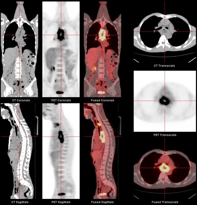
Positron-emission tomography (PET) is medical diagnostic technology that has been developed for over two decades and has become a clinical diagnostic tool for cancer and some cardiac diseases and neuropsychiatric disorders.
The division conducts more than 10,000 nuclear medicine imaging scans every year, including PET scans.

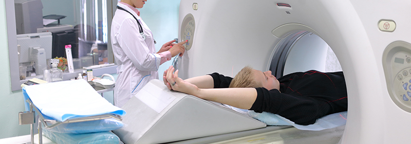
A noninvasive nuclear medical examination, PET injects radioisotopic tracers into the body via veins, and after they are fully absorbed by the cells in the body for metabolism, the radioactive rays emitted from inside the body are collected by the scanner and processed by computer to constitute images serving as a framework for clinical diagnosis.
PET examinations can detect lesions and contribute to early detection of the disease when functional abnormalities occur in cells and organs. We can also use some special tracers containing isotopes to treat cancer with their higher and longer binding force with the tumor.
PET can provide a variety of information about the disease for clinicians to complete the treatment scheme, as well as monitor the effect of therapy and surgery, which is of great help for prognosis follow-up.


▲
